Callistemon citrinus and Cistus salvifolius, Two New Hosts of Phytophthora taxon niederhauserii in Italy
S. O. Cacciola, S. Scibetta, A. Pane, R. Faedda, and C. Rizza.
October 2009
Bottlebrush (Callistemon citrinus (Curtis.) Skeels., Myrtaceae) and rock rose (Cistus salvifolius L., Cistaceae) are evergreen shrubs native to Australia and the Mediterranean Region, respectively. In the spring of 2003, approximately 2% of a nursery stock of 12-month-old potted plants of C. citrinus and 8% of a nursery stock of 12-month-old potted plants of Cistus salvifolius grown in the same nursery in Sicily, showed symptoms of leaf chlorosis, defoliation, and wilt associated with root and collar rot. A Phytophthora species was consistently isolated from roots and basal stems on BNPRAH selective medium (2). One isolate from rock rose (IMI 391708) and one from bottlebrush (IMI 391712) were characterized. On potato dextrose agar (PDA), the colonies showed stoloniform mycelium and irregular margins; on V8 juice agar (V8A), colonies were stellate to radiate. Minimum and maximum temperatures on PDA were 10 and 35°C, respectively, with the optimum at 30°C. Mean radial growth rate of isolates on this substrate was 9.9 and 11.3 mm/day, respectively. In saline solution (1), both isolates produced catenulate hyphal swellings and ellipsoid, nonpapillate, persistent sporangia with internal proliferations and dimensions of 52 to 70 × 30 to 42 µm and 51 to 85 × 39 to 45 µm. Mean l/b ratio of sporangia for both isolates was 1.8 ± 1. On V8A plus ß-sytosterol, both isolates produced amphyginous antheridia and spherical oogonia in dual cultures with an A2 tester of P. drechsleri Tucker. Conversely, they did not produce gametangia with an A1 tester of P. cryptogea Pethybr., indicating they were A1 mating type. The internal transcribed spacer (ITS)-rDNA sequences of rock rose and bottlebrush isolates showed 100% similarity with those of two reference isolates of P. taxon niederhauserii from GenBank (Accession Nos. FJ648808 and FJ648809). On the basis of the analysis of the DNA, the species isolated from bottlebrush and rock rose were identified as Phytophthora taxon niederhauserii. Pathogenicity tests were carried out on 6-month-old potted plants of C. salvifolius and C. citrinus (10 plants of each plant species for each isolate) transplanted into pots (12 cm in diameter) containing a mixture of 1:1 steam-sterilized, sandy loam soil (vol/vol) with 4% inoculum produced on autoclaved kernel seeds. Plants were maintained at 25 to 28°C and watered to soil saturation once a week. After 2 to 3 weeks, all inoculated plants developed symptoms identical to those observed on plants with natural infections. Ten control plants transplanted into pots containing noninfested soil remained healthy. P. taxon niederhauserii was reisolated solely from inoculated plants. To our knowledge, this is the first report of P. taxon niederhauserii on C. citrinus and C. salvifolius in Italy. This Phytophthora taxon has been reported recently on rock rose in Spain
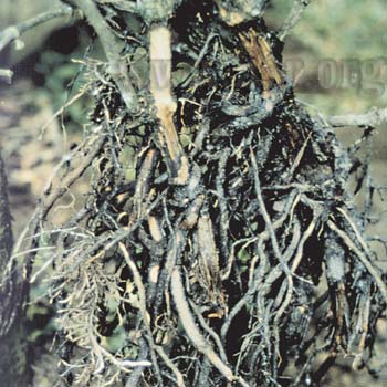
Root Rot of Hydroponically Grown Lettuce Caused by Phytophthora drechsleri in Mexico
G. Rodríguez-Alvarado, M. I. Pérez-Cáliz, K. B. Caudillo-Ruiz, E. Garay-Serrano, R. Rodríguez-Fernández, and S. P. Fernández-Pavía.
October 2009
During March of 2008, bibb lettuce (Lactuca sativa L.) plants with severe wilting and root rot were observed in a commercial liquid-hydroponic greenhouse in Guanajuato, Mexico. By July of that year, the disease affected most plants in the facility. A Phytophthora sp. was consistently isolated from diseased roots on potato carrot agar. Several Phytophthora isolates were morphologically characterized. Sporulation was achieved by placing colonized disks of clarified V8 juice agar (V8A) into nonautoclaved soil extract (10 g avocado soil/1,000 ml distilled water, stirred for 3 h, and filtered). Sporangia were persistent, nonpapillate, and 40 to 58 µm long × 30 to 40 µm wide. External and internal proliferation was observed. Hyphal swellings were predominantly rounded. Oospores were not observed. The isolates grew on V8A at 35°C. Pathogenicity tests were conducted twice by utilizing a representative isolate (AC1) on bibb lettuce seedlings (10 replicates per experiment). Seeds were placed on sterile, water-soaked paper in petri dishes. After 10 days, each lettuce seedling was placed into a tube containing approximately 2 ml of sterile distilled water and 2,000 zoospores. Control plants were placed in tubes with water only. Plants were incubated for 7 days in a moist chamber at 25°C. Symptoms of wilting and root necrosis were observed 2 to 3 days after inoculation. All plants were dead 5 to 7 days after inoculation. A Phytophthora sp. was always isolated from the roots of inoculated plants. Control plants remained healthy. The pathogen was identified as Phytophthora drechsleri Tucker according to morphological characteristics. To confirm the identity of the pathogen, sequences of the internal transcribed spacers (ITS) were obtained from three representative isolates. The ITS sequences that were obtained shared 100% homology to several strains of P. dreschleri, including isolates from cucurbits (GenBank Accession No. AF228097). The ITS sequence was deposited in NCBI as Accession No. FJ790770. P. cryptogea and P. dreschleri have been reported as causing root rot on lettuce grown hydroponically in the United States and Korea (1,2). To our knowledge, this is the first report of P. drechsleri causing root rot on lettuce in Mexico. Source:Plant Disease October 2009, Volume 93, Number 10
Page 1077
First Report of Root Rot and Twigs Wilting of Olive Trees in Argentina Caused by Phytophthora nicotianae
A. M. Vettraino, G. Lucero, P. Pizzuolo, S. Franceschini and A. Vannini
July 2009
In Argentina, olives (Olea europaea) are planted on approximately 90,000 ha located primarily in the northwest continental regions. During a 2005 survey, root rot was recorded at several olive plantations in Catamarca, La Rioja, and San Juan provinces (3). Aboveground symptoms associated with root rot were twigs wilting with or without chlorosis, defoliation, and death. Symptoms were initiated on lateral branches and sometimes affected the entire crown. Even if young (5-year-old) trees displayed root rot, aerial symptoms may or may not be seen until years later. Disease incidence varied from 3 to 30%. Rotted rootlets were associated mainly with the infection of Phytophthora palmivora Butler and less frequently with another Phytophthora species. Isolates of this species were heterothallic, had a fluffy growth on carrot agar, and arachnoid growth on potato dextrose agar. Chlamydospores approximately 36 µm in diameter were also produced. The species developed prominent, papillate, noncaducous sporangia of different shapes ranging from ellipsoid to spherical when submerged in saline solution. Sporangia were 35 to 57 × 25 to 45 µm (average 44 × 33 µm), L:B ratio from 1.1 to 1.7. Isolates formed oogonia and amphyginous antheridia following mating type assays. On the basis of morphological features, these isolates were identified as P. nicotianae Breda de Haan. Identity was confirmed by sequencing the rDNA internal transcribed spacer (GenBank Accession No. FJ746693) (1). One-year-old O. europea seedlings were challenged with P. nicotianae (A1 isolates 306G and 339) through soil infestation assay in a growth chamber at 25°C. Infested and uninfested autoclaved millet grains moistened with V8 juice were used to inoculate 15 olive seedlings per isolate and controls, respectively. Fifty days after inoculation, seedlings showed foliar symptoms similar to those observed in the field and had an average of 50% reduction in the root system. Control plants remained healthy. P. nicotianae was always reisolated from symptomatic roots. P. nicotianae was reported on Citrus aurantium in Argentina in 1947 and is currently associated with several hosts (2). In 2002, the same species was reported associated with olive root rot in southern Italy (4). It is possible that P. nicotianae was recently introduced into Argentina through importation of Mediterranean olive varieties. The demonstrated pathogenicity of P. nicotianae on olive together with the recently reported detection of P. palmivora (3) presents a serious threat to olive cultivation in Argentina. Source:Plant Disease uly 2009, Volume 93, Number 7
Page 765
First report of Phytophthora nicotianae causing black shank of Schizonepeta tenuifolia in China
Z. Zhang, J.-H. Wei*, L. Cao, C.-M. Yang and C Sui
Institute of Medicinal Plant Development, Chinese Academy of Medical Sciences and Peking Union Medical College, Beijing 100193, P.R. China
Jan 2009
Schizonepeta tenuifolia (Lamiaceae) is a medicinal herb widely planted as an economically important plant in China. During the summer of 2008, plants with symptoms of black shank disease were observed in many fields of the municipalities of Beijing and Hebei provinces. Initially black-brown lesions were observed on the stems and roots, and then affected plants eventually wilted and died without dropping leaves. A survey found that more than 80% of S. tenuifolia fields were affected, resulting in serious economic loss.
Diseased stem and root samples were surface disinfected in 1% NaOCl for one minute and transferred to oomycete selective medium (Masago et al., 1977) and incubated at 22°C for five days. Single spore cultures were obtained and initially identified as Phytophthora nicotianae based on morphological and physiological features. (Stamps et al., 1990; Erwin & Ribeiro, 1996). Genomic DNA was extracted from the purified cultures using Nucleospin Plant Kit (Macherey-Nagel Inc., Düren, Germany). The internal transcribed spacer (ITS) region of rDNA was amplified using primers ITS4 and ITS6. The amplicons were sequencedand showed 100% nucleotide and 100% amino acid sequence identity with the P. nicotianae reference sequence (844bp, GenBank Accession No. FJ215687).
Koch's postulates were satisfied by dipping plants roots into the spore suspension (105spores/ml) obtained from 10-day-old culture plates. Sterilized water was used for control plants. The inoculated plants were placed outside under natural environmental conditions (mean temperature 20°C). After 10 days, black-brown lesions were observed on the root and stem samples. P. nicotianae was reisolated from the lesions fulfilling Koch's postulates. No symptoms were observed on the control plants. To our knowledge, this is the first report of black shank disease onS. tenuifolia caused by P. nicotianae in China.
First Report of the A2 Mating Type of Phytophthora capsici Infecting Peppers (Capsicum annuum) in Taiwan
Z. M. Sheu, J. R. Chen, and T. C. Wang, AVRDC-The World Vegetable Center, P.O. Box 42, Shanhua, Tainan 74199, Taiwan, Republic of China
May 2009
Phytophthora capsici Leonion was first identified on pepper (Capsicum annuum L) in Taiwan in 1976. At that time, only the A1 mating type was present (2). In 2007, the A2 mating type of P. capsici was identified on tomato and eggplant in the central part of the country (1). During an excessively rainy period in mid-2008, many chili and sweet pepper fields in Taiwan suffered severe losses due to P. capsici. Symptoms included a foliar blight and stem, root, and fruit rot. Plants eventually wilted and died. Symptomatic plants were collected from chili- and sweet pepper-production areas in central, southern, and eastern Taiwan. Fifty-three isolates from single zoospores were identified by PCR using species-specific primers CAPFW/CAPRV2 (4). Mating type was determined by co-inoculating rape seed agar plates (3) with mycelial plugs of the tester and a known isolate. Pc134, maintained by the mycology unit at The World Vegetable Center, and 27220, provided by P. J. Ann at the Taiwan Agricultural Research Institute, were used as reference isolates of A1 and A2 mating types, respectively. Plates were examined microscopically for oospores after 5 to 7 days of incubation at 24°C in the dark. Of the 53 isolates, 15 were identified as the A2 mating type and the remaining 38 isolates were identified as A1. The A2 mating type was found in the central and southern regions while the A1 mating type was widely distributed across all three regions. The sporangia of the A2 mating type were 60.4 to 73.4 × 40.9 to 51.8 ?m (average 69.2 × 44.7 ?m), whereas sporangia of the A1 mating type were 50.1 to 73.9 × 37.9 to 48.1 ?m (average 61.4 × 44.1 ?m). In general, the A2 mating type produced longer sporangia and only a few isolates produced chlamydospores in V8 broth and on agar. To our knowledge, this is the first report of the A2 mating type of P. capsici infecting peppers in Taiwan. The presence of both mating types in the same field has been observed.
First Report of Phytophthora citricola on Cornus mas in Bulgaria
S. G. Bobev, K. Van Poucke and M. Maes.
Institute for Agricultural and Fisheries Research, Plant-Crop Protection, 9820 Merelbeke, Belgium
May 2009
Cornelian cherry dogwood (Cornus mas) is a widespread species in Bulgaria and some cultivars with large fruits are the subject of propagation. In the springs of 2007 and 2008, severe, unusual damages were observed on sporadically scattered plantlets of 'Kazanlashki' (known also as 'Kazanlaker') in a nursery located near Vratza in northwestern Bulgaria. Symptoms were identical in both years and expressed on the leaves, young shoots, and adjacent rootstock wood. Dark brown, necrotic leaf spots initiated most often from the leaf periphery and quickly covered more than half of the leaf area. Necrosis of the leaves and shoots spread toward the older woody tissues and the plantlets died within a couple of weeks. Isolations from symptomatic leaves, shoots, and rootstocks (three to five samples per plant organ) on potato dextrose agar always revealed a fungus-like organism that formed relatively fast-growing white, radial, petaloid colonies. Numerous, ovoid to obpyriform, noncaducous, semipapillate sporangia occasionally with two papilla were observed after 1 or 2 days of incubation at 20°C in nonsterile soil extract (1). Average sporangium size was 39 (35 to 45) × 31 (20 to 35) ?m with a ratio between both parameters of approximately 1.26. The pathogen's paragynous antheridia and smooth-walled spherical oogonia (20 to 32 ?m in diameter) yielded spherical aplerotic to almost plerotic oospores on V8 medium with an average size of 25 ?m. The morphological data identified the organism as Phytophthora citricola (1). Isolates had identical cultural and morphological characteristics, and pathogenicity was tested by laboratory inoculations carried out in 2007 (two isolates) and twice in 2008 (three isolates). Separately, detached leaves of C. mas seedlings and 'Kazanlashki' were wiped with 70% ethanol, punctured with a needle, and the wounds inoculated with 5-mm mycelial plugs from a 7-day-old V8 growth plate. Sterile V8 plugs were placed onto similar wounds of control leaves. Leaf samples were incubated at 20°C in a humidified chamber. Necrosis similar to that observed in the field became visible around the mycelia plugs 4 days after inoculation. The necrotic lesions enlarged to 20 to 25 mm in diameter within the next 2 days, whereas the control leaves did not show any symptoms. Subsequently, the pathogen was reisolated solely from all the mycelium-inoculated samples. By means of the same inoculation method, pathogenicity was also demonstrated on shoots and mature fruits of C. mas. DNA was isolated from mycelium of an isolate and the internal transcribed spacer (ITS) region was amplified using ITS6 and ITS4 primers. The PCR product was sequenced (GenBank Accession No. FJ269034) and the BLAST search showed 100% homology with P. citricola, type II (2). To our knowledge, this is the first report of P. citricola on C. mas in Bulgaria, thus confirming its ability to attack Cornus spp. (3). Taking the lethal results of the disease and the polyphagous nature of the causal agent into consideration, this report is a serious warning for nurserymen and consumers.
Collar Rot on Italian Alder Trees in California Caused by Phytophthora siskiyouensis
S. Rooney-Latham, C. L. Blomquist, T. Pastalka, and L. Costello
April 2009
In November 2006, trees of Italian alder were observed declining in association with bleeding trunk cankers in a commercial landscape in Foster City, CA. A species of Phytophthora was isolated on PARP selective medium from the leading edge of the cankers. The Phytophthora species was homothallic with primarily paragynous antheridia and had oospores that were mostly globose and aplerotic. Sporangia were produced from mycelia on plugs of carrot piece agar in soil extraction solution and were semi-papillate and ovoid to ellipsoid in shape. The intergenic transcribed spacer region of rDNA from an alder isolate matched with 100% identity to isolates in GenBank of Phytophthora siskiyouensis. Pathogenicity was confirmed on Italian alder trees and potential pathogenicity was demonstrated on red and white alder trees.
Acknowledgements
Plant Disease
The British Society for Plant Pathology- New Disease Reports.